For further information regarding the INR, please see the following sources, accessible by clicking on the links below:
Comments: 0
Intracerebral hemorrhage (ICH) is a devastating stroke subtype characterized by a prominent neuroinflammatory response. Antagonism of pro-inflammatory cytokines by specific antibodies represents a compelling therapeutic strategy to improve neurological outcome in patients after ICH. To test this hypothesis, the tumor necrosis factor alpha (TNF-alpha) antibody CNTO5048 was administered to mice after ICH induction, and histological and functional endpoints were assessed.
Using 10 to 12-week-old C57BL/6J male mice, ICH was induced by collagenase injection into the left basal ganglia. Brain TNF-alpha concentration, microglia activation/macrophage recruitment, hematoma volume, cerebral edema, and rotorod latency were assessed in mice treated with the TNF-alpha antibody, CNTO5048, or vehicle.
After ICH induction, mice treated with CNTO5048 demonstrated reduction in microglial activation/macrophage recruitment compared to vehicle-treated animals, as assessed by unbiased stereology (P = 0.049). This reduction in F4/80-positive cells was associated with a reduction in cleaved caspase-3 (P = 0.046) and cerebral edema (P = 0.026) despite similar hematoma volumes, when compared to mice treated with vehicle control. Treatment with CNTO5048 after ICH induction was associated with a reduction in functional deficit when compared to mice treated with vehicle control, as assessed by rotorod latencies (P = 0.024).
Post-injury treatment with the TNF-alpha antibody CNTO5048 results in less neuroinflammation and improved functional outcomes in a murine model of ICH.
Comments: 0
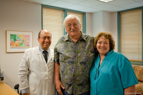
Charlie and Cheryll Giles with Dr. Tobinick at the INR 100 UCLA Medical Plaza July 11, 2013
Three years after the original filming of “A New Shot at Life” by 60 Minutes Australia.
To view the documentary, please go to the original 60 Minutes Australia feature, available here.
Comments: 0
On March 30, 2013, new data from the UCSF Memory and Aging Center published pointing to excess TNF as a potential therapeutic target for certain forms of dementia. In particular, elevated TNF levels were observed in the cohort of individuals examined with semantic variant PPA (primary progressive aphasia) (see Miller ZA, Rankin KP, Graff-Radford NR, Takada LT, Sturm VE, Cleveland CM, Criswell LA, Jaeger PA, Stan T, Heggeli KA, et al: TDP-43 frontotemporal lobar degeneration and autoimmune disease. J Neurol Neurosurg Psychiatry 2013).
We note that this UCSF study follows by more than four years the initial report of rapid clinical improvement in an individual with PPA following the off-label use of perispinal etanercept (Tobinick E: Perispinal etanercept produces rapid improvement in primary progressive aphasia: identification of a novel, rapidly reversible TNF-mediated pathophysiologic mechanism. Medscape J Med 2008, 10:135).
Comments: 0
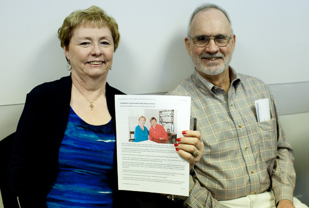
We were so pleased that Jan returned to visit us on March 11 from Kennett, Missouri with her husband. She is holding the story the Daily Dunklin Democrat had printed on February 17 about her rapid improvement after treatment. To read the whole story please click on the following link to the webpage of the Daily Dunklin Democrat (click here).
Comments: 0